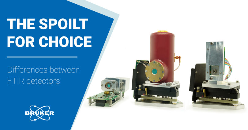
Do you feel the spolit for choice in choosing the right FTIR detector for your application? With this blog article, we would like to help you with this endeavor. We will discuss:
- why choosing the right detector is so important in FTIR microscopy
- different FTIR detectors and their advantages and disadvantages
- which detectors are suitable for your application
Challenges in IR microscopy
FTIR microscopy is a powerful tool. It combines conventional light microscopy with chemical identification by FTIR spectroscopy. This technique allows us to measure samples below 300 µm up to single-digit micrometers.
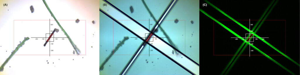
In IR microscopy objective lenses with a fixed field of view are used. An aperture is required to make sure that only relevant chemical information reaches the detector. It is placed over the structure to be investigated. Depending on the sample size, only a small amount of IR radiation can reach the detector. Because of this, highly sensitive detectors are needed to generate interpretable IR spectra.
Single-element FTIR detectors
In principle, two different single-element detectors are used in FTIR microscopy:
DTGS detectors (Deuterated alanine doped Tri-Glycine Sulphate) are based on a pyroelectric effect. Here, incoming IR radiation heats and expands the detector material. They are versatile and don’t need to be cooled by liquid Nitrogen. Additionally, this FTIR detector works particularly well under room temperature conditions. However, they require a high-amount of IR radiation to produce spectra of decent quality.
MCT detectors (Mercury cadmium telluride) are based on an internal photoelectric effect. Here, incoming IR radiation excites electrons into the conduction band. This excitation can also be triggered by thermal processes. Thus, it is necessary to cool MCT detectors to suppress thermally induced noise. As a result, they offer highest sensitivity in IR applications. They are typically used in low-light scenarios (e.g. protein analysis in water or microscopy). MCT FTIR detectors offer significantly better S/N ratios and exhibit much faster response times than DTGS detectors.
Traditionally, liquid nitrogen is used for the cooling (LN2-MCT). Unfortunately, the refilling of the nitrogen during prolonged use and cooling down takes time and creates additional costs. However, in recent years, cryogen-free, thermoelectrically cooled (TE-) MCTs have emerged. These detectors increase sensitivity at low maintenance.
Cryogen-free FTIR detector comparison
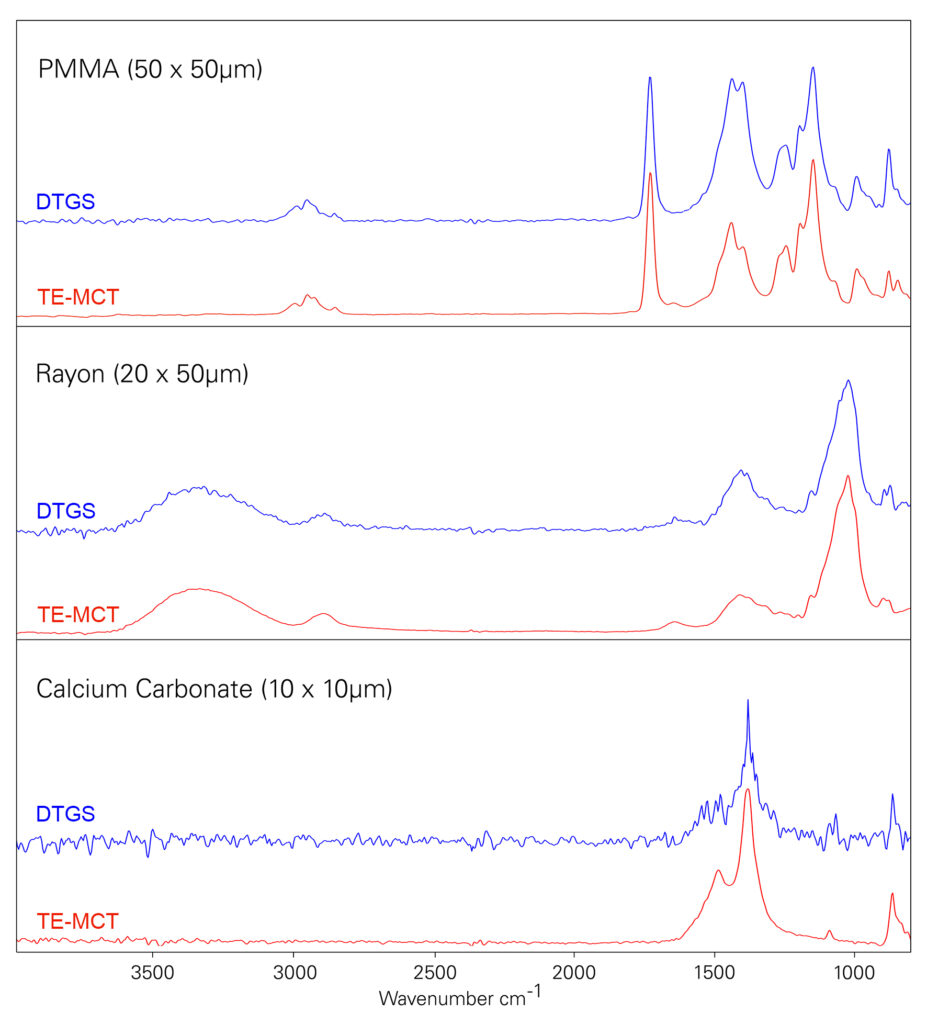
We wanted to emphasize the difference between the detectors. Therefore, IR spectra of polymer particles of different sizes were acquired. To enable a fair comparison, all measurements were performed under the same conditions.
In all three examples the DTGS detector shows considerable noise. For PMMA (50 x 50 µm) the spectral quality is still debatable. The FTIR spectra of Rayon and calcium carbonate show problematic artefacts. This can easily lead to misinterpretation. Thus, the DTGS disqualifies for use in FTIR microscopy below 50 µm.
The TE-MCT on the other hand outclasses the DTGS in every way. All spectral features in all three examples are precisely resolved, even at a 10 x 10 µm aperture. Consequently the TE-MCT detector gives access to the single-digit micrometer range in FTIR microscopy. Thus, the TE-MCT detector is the device of choice when it comes to cryogen-free detectors.
MCT FTIR detector comparison
Let’s put the performance of TE-MCT detectors into perspective. We compared it against liquid nitrogen cooled MCT detectors. The experimental setup was identical to the comparison between DTGS and TE-MCT detectors. Our results are displayed below.
The LN2-MCT detector shows a better S/N ratio compared to the TE-MCT detector. However, the differences only get noticeable as we enter high resolution territory. For 50 x 50 µm and 20 x 50 µm apertures spectra generated by the TE-MCT are almost indistinguishable.
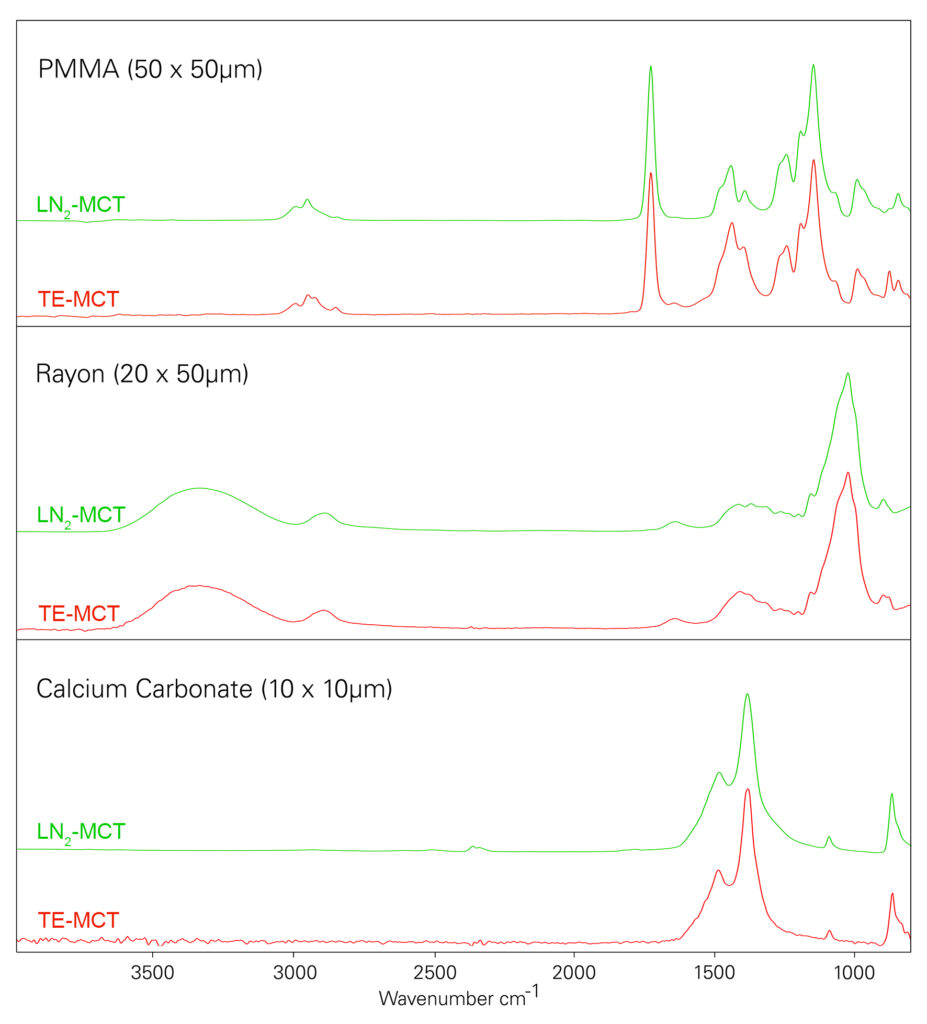
TE-MCT detectors achieve the long sought-after balance of good spectral quality and cryogen-free operation. If you are interested in convenient routine high-quality measurements the TE-MCT is perfect. No liquid nitrogen, no cool-down, always ready.
However, LN2-MCT detectors offer a reasonable advantage in applications within the single-digit micrometer range. These include scientific or forensic applications.
Conclusions and a clear winner
- Cryogen-free TE-MCT outperforms DTGS detectors
- An LN2-MCT offers better performance <10 µm
All our data shows, what the optimal configuration for an FT-IR microscope is: a TE-MCT for routine single-point measurements